
Figure 3.
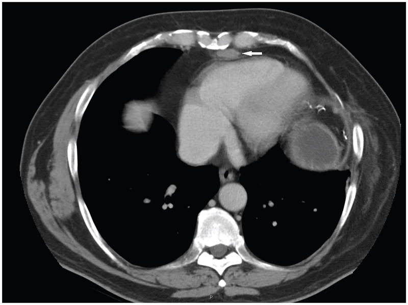
CT of the chest of an MEN1 patient with a history of thymic neuroendocrine tumor and prior surgical resection, showed a 2.5 × 1.2 cm suspicious mediastinal lesion (marked by arrow). It was resected and proven to be a thymic neuroendocrine tumor by surgical pathology.