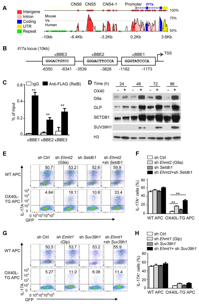
Figure 5. RelB recruits histone methyltransferases G9a and SETDB1 to the IL-17 locus.
(A) Vista analysis showing CNS regions at Il17a locus (mouse vs. human). Positions are assigned relative to the transcriptional start site (TSS) of mouse Il17a (horizontal axis).
(B) The consensus sequences and positions of NF-κB binding sites in the region of 10 kb upstream of mouse Il17a, numbers below the diagram indicate relative position to the transcriptional start site (TSS).
(C) ChIP analysis of the enrichment of RelB at the Il17a locus in WT CD4+ T cells transduced with retrovirus expressing FLAG-RelB and cultured for 24 h under Th17-polarizing conditions. ** p <0.01
(D) Immunoblot analysis of the expression of G9a, GLP, SETDB1, and SUV39H1 in the nucleus of WT CD4+ T cells activated as in Figure 2B. Data are representative of three independent experiments.
(E and F) Induction of Th17 cells from WT CD4+ T cells transduced with empty vector (sh Ctrl) or with retroviruses expressing shRNA oligos targeting Ehmt2 (G9a), Setdb1 and cultured for 3 d under Th17-polarizing conditions (E). Graphs in (F) depict Mean ± SD of 3 experiments. ** p <0.01
(G and H) Induction of Th17 cells from WT CD4+ T cells transduced with empty vector (sh Ctrl) or with retroviruses expressing shRNA oligos targeting Ehmt1 (Glp), Suv39h1 and cultured for 3 d under Th17-polarizing conditions (G). Graphs in (H) depict Mean ± SD of 3 experiments. ** p <0.01

