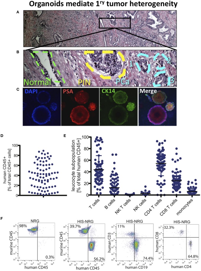
Figure 2.
PCa organoids to model tumor heterogeneity and develop immunotherapy in humanized mice. (A) H&E of RP section from a PCa patient shown in 4x. (B) The outlined area in (A) is displayed in 200x, showing the outline of benign prostate gland (green), prostatic intraepithelial neoplasia (PIN) region (yellow), and a three foci region of PCa (Blue). (C) Single-cell organoids reflect the heterogeneity in primary PCa. Immunofluorescence (IF) images show DAPI as nuclear staining, PSA (center region), CK14 (in cells lacking PSA staining, i.e., transit amplifying cells). Multiple organoids derived from the same patient's PCa expressing PSA and CK14 (right), low and localized (top) and low/negative (bottom). (D) Human immune system (HIS) reconstitution in NRG HIS. Fraction of human CD45+ cells of total CD45+ cells detected in the HSC transplanted NRG mice. (E) Indicated leukocyte subpopulations were determined by FACS analysis of PBMC in NRG mice. (F) Indicated immune cell subpopulations in NRG and HIS-NRG mice are shown as dot plots.

