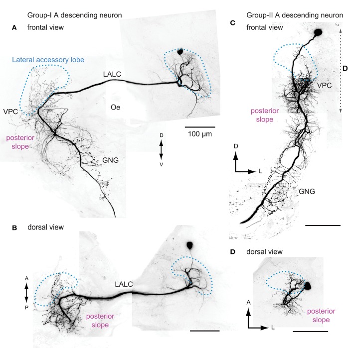
Figure 4.
The morphology of flip-flop descending neurons innervating the lateral accessory lobe in the silkmoth. Frontal and dorsal views of the group-IA DN (A,B) and the group-IIA DN (C,D) are shown. Group-IA DN has smooth processes mainly within the ipsilateral LAL and varicose processes in the contralateral LAL, ventral protocerebrum, posterior slope, and the gnathal ganglia (A,B). Group-IIA DN has smooth processes in the ipsilateral LAL, ventral protocerebrum and posterior slope, and varicose processes in the ipsilateral posterior slope, gnathal ganglia (C,D). Outline of the LAL is shown with broken line (blue). The range of maximum intensity projection for (D) is shown by broken line in (C). GNG, gnathal ganglion; Oe, esophagus; VPC, ventral protocerebram. Images are prepared based on the data used in Mishima and Kanzaki (1999). A, anterior; LALC, lateral accessory lobe commissure; GNG, gnathal ganglia; Oe, esophagus; P, posterior; VPC, ventral lateral protocerebrum.

