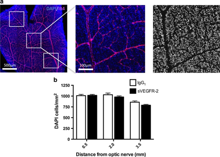
Figure 5.
Reductions in CTB transport precede cell body loss. (a) Retinas from 48- h post-CTB injection transport experiments were stained with DAPI (blue) and IB4 (magenta) (left panel). Tile scan images of each of the four petals were taken (original magnification= × 10), 0.5mm2 areas were selected at 0.5, 2.0 and 3.5mm from the optic nerve (middle panel), and then processed to remove the vascular area from each picture (right panel). DAPI-stained nuclei were counted using ImageJ software. (b) A comparison of DAPI nuclei per mm2 revealed no significant differences between IgG1 or sVEGFR-2 treatments at 0.5, 2.0 or 3.5mm from the optic nerve. These data indicate that at 1-week post-sVEGFR-2 injection, transport deficits may precede significant RGC loss. N=5–6 retinas. Data are given as means±S.E.M.

