Figure 5.
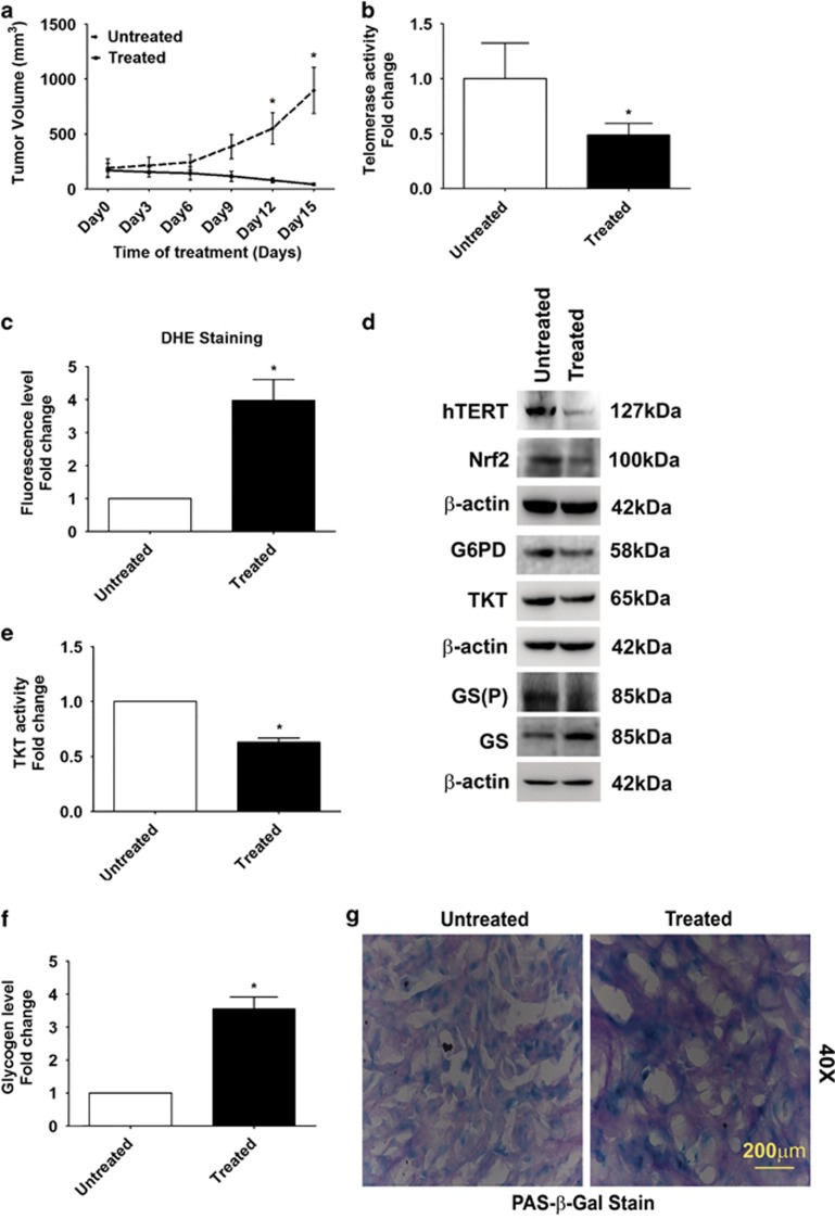
Costunolide inhibits pentose phosphate pathway and increases glycogen accumulation in heterotypic xenograft glioma model. (a) Significant reduction in tumor volume in Costunolide-treated glioma xenografts as compared with untreated groups (n=7). (b) Costunolide-treated tumors show decreased telomerase activity as compared with control groups (n=4). (c) Elevated ROS levels in Costunolide-treated tumors as compared with untreated group, as indicated by increased DHE fluorescence (n=4). (d) Western blot showing decreased hTERT, Nrf2, G6PD, TKT, and GS (P) levels in the total cell lysates prepared from Costunolide-treated tumors as compared with untreated groups. Blot is representative images (n=6). Blot was re-probed for β-actin to establish equivalent loading. (e) Decreased TKT activity and (f) elevated glycogen levels in Costunolide-treated tumors as compared with untreated controls. The values in graph (b, c, e, and f) indicate means±S.E.M. of n=4. Statistical analysis was done using unpaired Student's t-test. Results were considered as significant, when P-value was equal to or less than 0.05. *Denotes significant change from the untreated group. (g) Costunolide increases glycogen accumulation in cells undergoing senescence as demonstrated by increased PAS and β-glactosidase staining. Sections derived from Costunolide-treated U87 xenografts were co-stained for β-glactosidase and glycogen (PAS). Representative image from animals from each group is shown (n=4)

