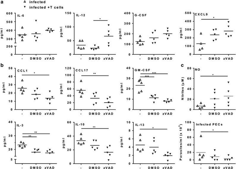
Figure 8.
Inhibition of T-cell apoptosis enhances macrophage-mediated immunity to T. cruzi infection. (a–c) Splenic T cells from infected mice (18 dpi) were treated with anti-CD3 in the presence of caspase inhibitor zVAD or dimethyl sulfoxide (DMSO) for 4 h. T cells were washed and adoptively transferred intraperitoneally to infected mice (20 dpi). Infected mice injected with phosphate-buffered saline (PBS) only were used as controls. After 2 days, peritoneal exudates were analyzed for (a) M1 and for (b) M2 cytokines. (c) Peritoneal macrophages were cultured during 24 h before evaluation of spontaneous NO production or infected with T. cruzi and cultured during 4 weeks before determination of parasite burden. (a and b) Symbols represent peritoneal exudates from individual mice injected with PBS (Δ) or with activated T cells (▾) previously treated with zVAD or DMSO (N=5 mice per group). In (c), each symbol represents the mean of two to three technical replicates of cultured cells from each individual mouse. Significant differences between treatments are indicated (*), as analyzed by t-test (c) or by analysis of variance (ANOVA) with Tukey's post-test (a and b)

