Figure 6.
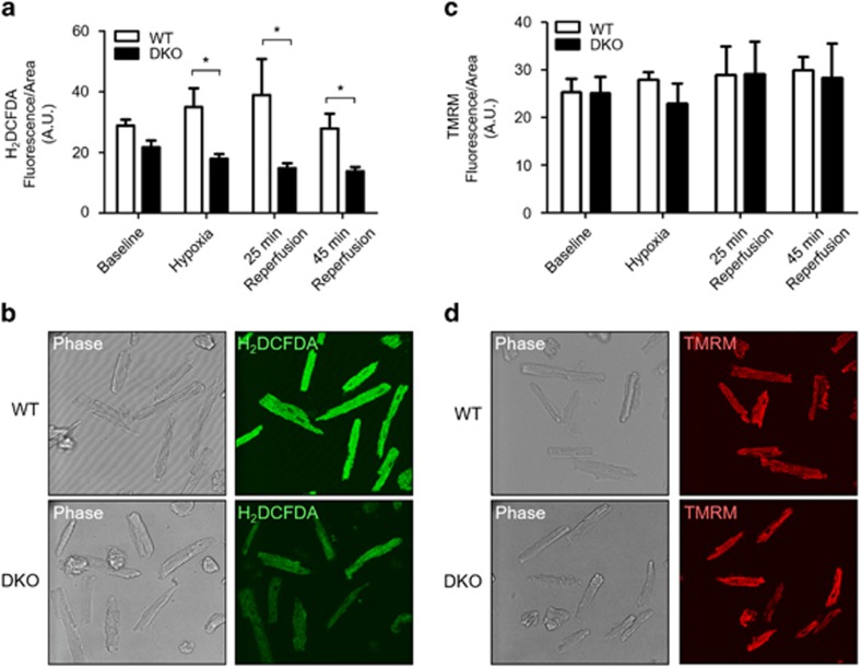
Acute ablation of both cardiac Mfn1 and Mfn2 attenuated production of oxidative stress during acute I/R injury. (a) The increase in oxidative stress (measured by H2DCFDA fluorescence) observed in WT cardiomyocytes during simulated ischemia and reperfusion was attenuated in DKO cardiomyocytes. There was a statistical difference in oxidative stress handling between the two genotypes across the I/R protocol (P<0.001, two-way ANOVA), with further statistical significance at 25 min after simulated reperfusion (Bonferroni post-test P<0.01). (b) Representative phase contrast and confocal H2DCFDA images of WT and DKO cardiomyocytes taken at the end of simulated ischemia. (c) There was no difference in mitochondrial membrane potential (measured by 30 nM TMRM fluorescence) under baseline conditions, at the end of simulated ischemia and 25 and 45 min after simulated reperfusion, between DKO and WT ventricular cardiomyocytes (two-way ANOVA with Bonferroni post-test). (d) Representative phase contrast and confocal and TMRM images of WT and DKO cardiomyocytes taken at the end of simulated ischemia. N=4 mice/group. Error bars are S.E.M and statistical analysis was performed by a two-way ANOVA. *P≤0.05

