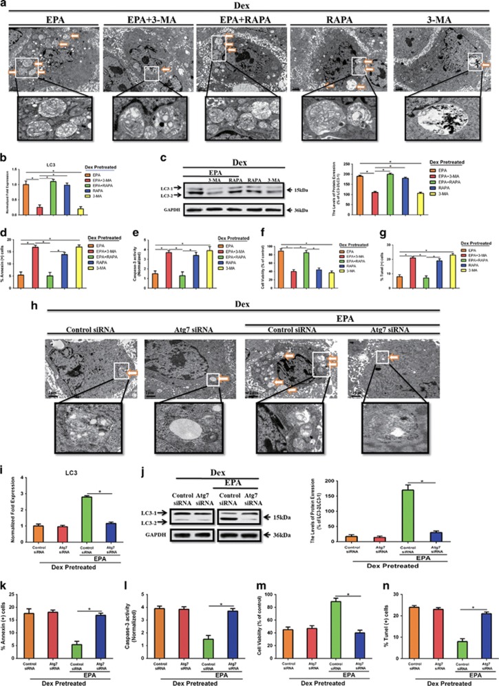
Figure 2.
EPA inhibited Dex-induced apoptotic cell death via inducing adaptive cell autophagy. mBMMSCs were cultured with or without 10−6 M Dex and 100 μM EPA for 24 h in the presence or absence of 100 nM rapamycin (RAPA) and 5 mM 3-methyladenine (3-MA). (a) Fixed cells processed for thin-section electron microscopy. Arrows indicate the autophagic vacuoles digesting organelles or cytosolic contents (magnification, × 2500). (b) RT-PCR analysis of LC3 treated with certain groups. (c) Western blot analysis and quantification of LC3 (LC3-2/LC3-1) protein treated with certain groups. (d) Annexin V/PI double staining was performed to detect apoptotic cells. (e) Caspase-3 activity was detected by the caspase-3 assay kit in mBMMSCs that were treated with or without 10−6 M Dex and 100 μM EPA for 24 h in the presence or absence of 100 nM rapamycin (RAPA) and 5 mM 3-methyladenine (3-MA). (f) The cultures were incubated with or without Dex (10−6 M) and 100 μM EPA for 24 h in the presence or absence of 100 nM rapamycin (RAPA) and 5 mM 3-methyladenine (3-MA). Cell viability was determined by MTT assay. (g) Cell apoptosis was detected by TUNEL assay and number of TUNEL-positive cells as a percentage of the positively stained mBMMSCs. Expression of each target gene was calculated as a relative expression to β-actin and represented as normalized fold expression. (h) Atg7 was knocked down by siRNA and cells were cultured with 10−6 M Dex with or without 100 μM EPA. Fixed cells processed for thin-section electron microscopy. Arrows indicate the autophagic vacuoles digesting organelles or cytosolic contents (magnification, × 2500). (i and j) RT-PCR, western blot analysis and quantification of LC3 (LC3-2/LC3-1) treated with certain groups. (k) Annexin V/PI double staining was performed to detect apoptotic cells. (l) Caspase-3 activity was detected by the caspase-3 assay kit in control or Atg7 knocked down mBMMSCs. (m) Cell viability was determined by MTT assay. (n) Cell apoptosis was detected by TUNEL assay and number of TUNEL-positive cells as a percentage of the positively stained mBMMSCs. The data are represented as the mean±S.D. of three independent experiments. *P<0.05 compared with each group (a–g). *P<0.05 compared with control siRNA treated with EPA (i–n). 3-MA, 3-methyladenine; RAPA, rapamycin

