Figure 2.
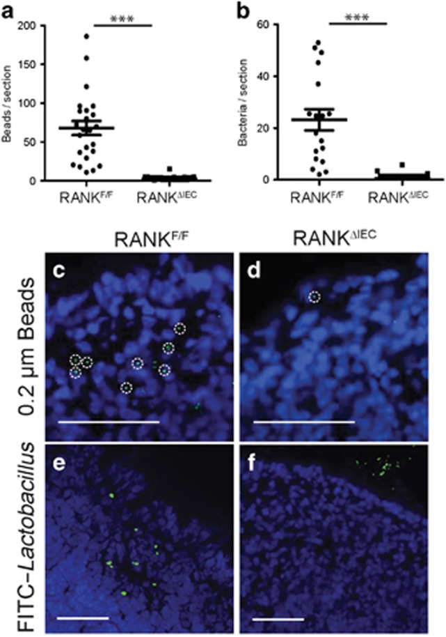
Peyer's patches (PPs) lacking M cells have reduced capacity to phagocytose particulate antigens. RANKF/F and RANKΔIEC mice were gavage fed with either 1 × 10¹¹ 0.2-μm diameter fluorescein isothiocyanate (FITC)-labeled polystyrene beads (a) or 1 × 10⁹ colony-forming unit (CFU) FITC-labeled L. rhamnosus strain GG (LGG) (b), followed by excision of the PPs after 6 h (a) or 24 h (b). Individual points on the scatter plots represent the number of beads or bacteria manually counted in cryosections of a single PP follicle (a) or the entire PP (b). (c,d) Representative images showing 0.2 μm diameter beads within PPs from RANKF/F (c) and RANKΔIEC (d) mice. The dashed circles show individual phagocytic cells containing multiple beads. (e,f) Representative images showing FITC-LGG within PPs from RANKF/F (e) and RANKΔIEC (f) mice. Bar = 100 μM. Data from each group are summarized as mean±s.e.m. and are representative of two experiments with three mice per group. ***P<0.001 (t-test).

