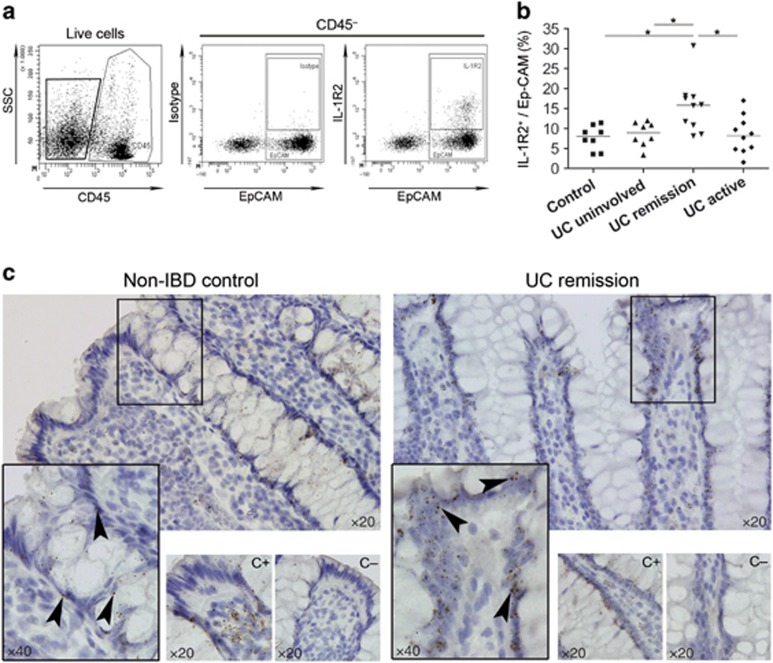
Figure 4.
Increased numbers of epithelial cells express IL-1R2 in UC patients in remission. (a) Representative flow cytometry dot plots from digested biopsies. (b) Dot plot representation (line as median) of the percentages of intracellular IL-1R2 staining among Ep-CAM+ from CD45− cells from controls (n=8), uninvolved mucosa from UC patients (n=8), mucosa from UC patients in remission (n=10), and active UC patients (n=10). Data were analyzed using a Kruskal–Wallis test, followed by a Benjamini–Hochberg post–hoc correction test. *P<0.05. (c) In situ hybridization of IL1R2 transcripts in colonic lamina propria from control and UC patient in remission. Sections were counterstained with hematoxylin. Images were taken with × 20 and × 40 objective lenses. Hs-PPIB probe as a positive control (C+) and DapB probe as a negative control (C−). IL, interleukin; UC, ulcerative colitis.

