
Figure 1.
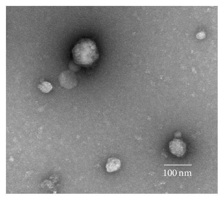
Transmission electron micrograph of a sample of the pellet obtained by ultracentrifugation of Coon's Modified Ham's F-12, 6-hormone medium used to grow FRTL-5 cells. Objects in the frame exhibit the characteristic shape of microvesicles under transmission electron microscopy, a spherical shape between 30 and 100 nm in diameter.