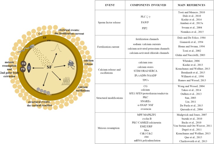
Figure 2.
Sperm-induced oocyte activation.
Left panel: image representing events occurring during sperm-induced oocyte activation: upon release of the SF, electrical modification of oocyte plasma membrane properties generates an outward (in mammals) or inward ion current (non-mammals). Release of calcium from the intracellular stores generates calcium oscillations. Physical changes of the oocyte occur by release of CG contents, and finally meiosis is resumed, allowing completion of the cell cycle, extrusion of the polar body and triggering of zygote formation. Right panel: table reporting the event, the components involved and relevant references. PLCζ, phospholipase C; PAWP, postacrosomal sheath WW domain-binding protein; PIP2, phosphatidylinositol (4,5)-bisphosphate; IP3, inositol 1,4,5-trisphosphate; cADPr, cyclic adenosine diphosphoribose; NAADP, nicotinic acid adenine dinucleotide phosphate; TPCs, two-pore channels; SFE1, SFE9, structural matrix proteins; PKC, protein kinase C; SNAP, N-ethylmaleimide-sensitive factor attachment protein alpha; NSF, N-ethilmaleimide sensitive factor; MPF, maturation promoting factor; MAPK, mitogen-activated protein kinase; CAMKII, calcium calmodulin-dependent protein kinase; EMI/ERP, early mitotic inhibitors; Mos, serine/threonine kinase; Cdk1, cyclin-dependent kinase; CG, cortical granule; SF, sperm factor.

