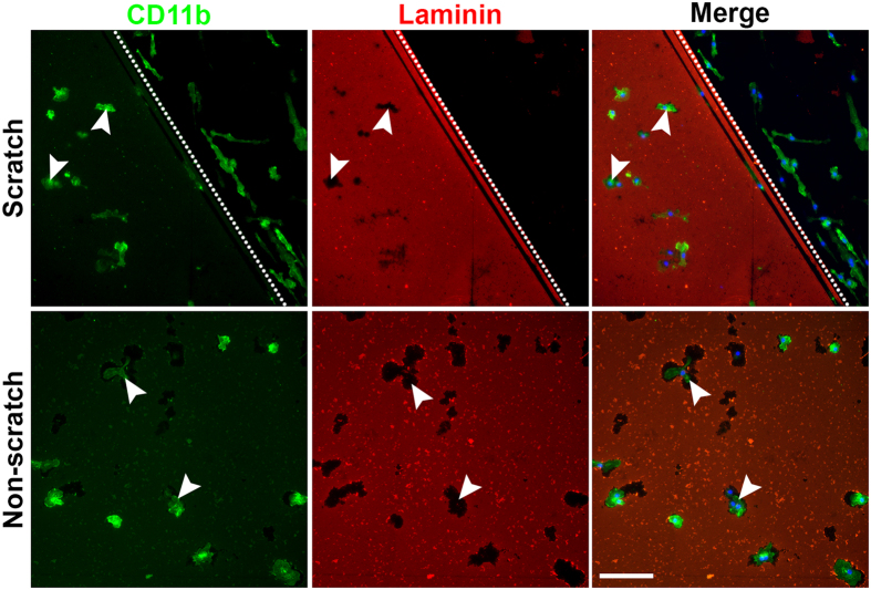
Figure 3. Amoeboid microglia digest the laminin coating, leaving laminin-free zones around the cells.
Amoeboid microglia (white arrowheads) digested the surrounding laminin on non-scratched areas of the PDL/laminin-coated surface, leaving some laminin-free zones. The bipolar/rod-shaped microglia colonized only on the scratched area. Scale bar: 100 μm.

