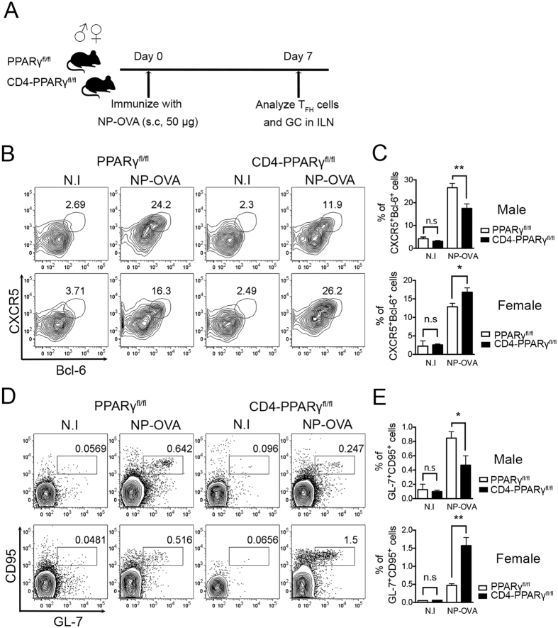
Figure 1. Female CD4-PPARγKO but not male mice have higher proportions of TFH cells and GC B cells.
(A) TFH cells were induced by immunization of six- to eight-week-old male and female littermate control and CD4-PPARγKO mice with NP-OVA. Seven days after immunization, the proportion of TFH cells was analyzed by flow cytometry. (B,C) NP-OVA-immunized male and female littermate control and CD4-PPARγKO mice were analyzed by staining with anti-Bcl-6 and anti-CXCR5 antibodies. Bcl-6 and CXCR5 double-positive cells gated on CD4+CD44high were identified as TFH cells and the % of Bcl-6+CXCR5+ TFH cells was represented as a bar graph. (D,E) Germinal center (GC) B cells in male and female littermate control and CD4-PPARγKO mice were examined by staining with anti-GL-7 and anti-CD95 antibodies seven days after NP-OVA immunization and the % of GL-7+CD95+ GC B cells gated on B220-positive cells was indicated as a bar graph. The data represent means ± SEM (n = 4/group, three independent experiments). *P < 0.05, **P < 0.01 by a two-tailed, unpaired Student’s t-test.

