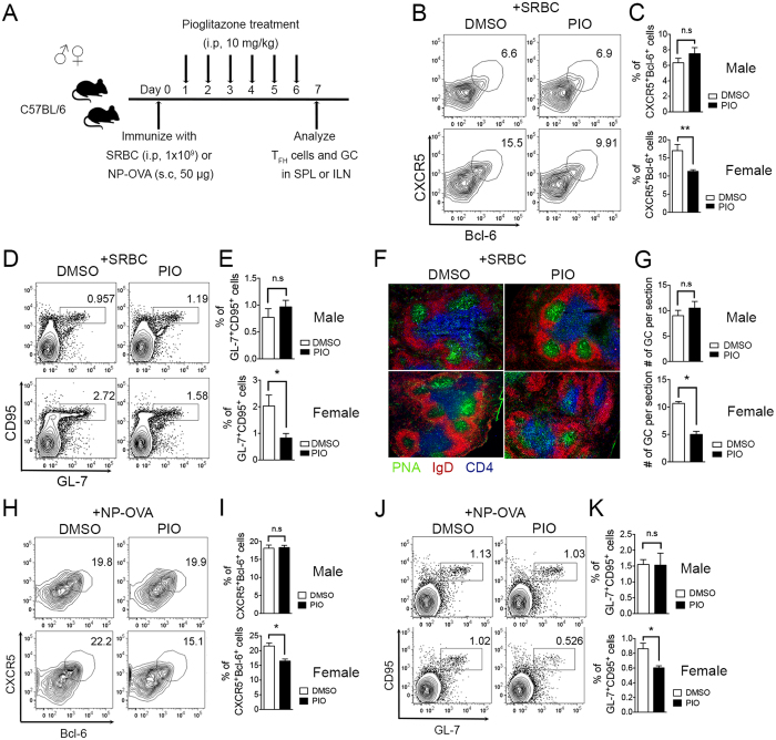
Figure 2. PPARγ ligand pioglitazone treatment reduces TFH responses in female but not in male mice.
(A) Six- to eight-week-old male and female mice were immunized with SRBC or NP-OVA and were treated daily with pioglitazone (10 mg/kg) intra-peritoneally from day 1 to day 6. (B,C) The mice were sacrificed following SRBC immunization and the spleens were isolated and stained with anti-mouse CXCR5 and Bcl-6 antibodies to analyze the proportion of TFH cells and the % of CXCR5+Bcl-6+-positive TFH cells was represented as a bar graph. (D,E) GL-7 and CD95 double-positive cells gated on B220-positive cells from the spleens from male and female mice were analyzed and the % of GL-7+CD95+ GC B cells was demonstrated as a bar graph (n = 3/group, three independent experiments). (F,G) Immunofluorescence analysis was performed to determine the number of GCs formed in spleens of SRBC-immunized male and female mice by staining with anti-PNA, -IgD, and -CD4 antibodies and the numbers of GCs were counted per spleen section. The average number of GCs per spleen section was determined (n = 9). (H,I) The inguinal lymph nodes were isolated from NP-OVA-immunized male and female mice. CXCR5+Bcl-6+-positive TFH cells were analyzed and displayed as a bar graph. (J,K) GL-7 and CD95 double-positive cells gated on B220-positive cells in the inguinal lymph nodes from male and female mice were analyzed and the % of GL-7+CD95+ GC B cells was represented as a bar graph. The data shown represent means ± SEM (n = 3 per group, two independent experiments). *P < 0.05, **P < 0.01 by a two-tailed, unpaired Student’s t-test.

