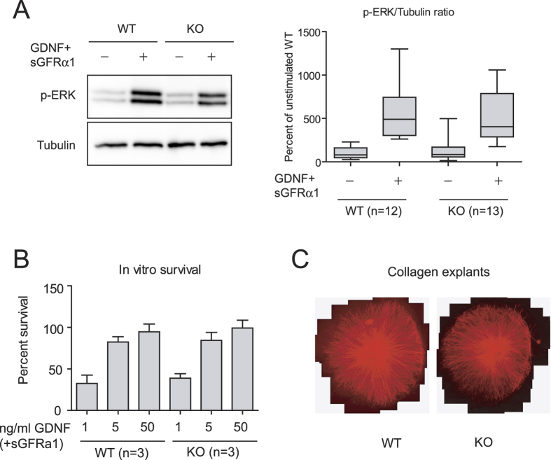
Figure 4. Dhcr7−/− sympathetic neurons properly respond to GDNF stimulation.
(A) Superior cervical ganglion neurons from wild type and Dhcr7 knockout were cultured and stimulated with GDNF as described in methods. Total lysates were resolved by SDS-PAGE and probed with the indicated antibodies (left panel). The graph on the right panel shows the relative intensity of phospho-ERK signal normalized by tubulin. (B) GDNF-mediated survival of SCG neurons was evaluated after 48 hours in the presence of growing concentrations of GDNF. (C) Representative images of SCG explants after five days in the presence of 50 ng/ml GDNF.

