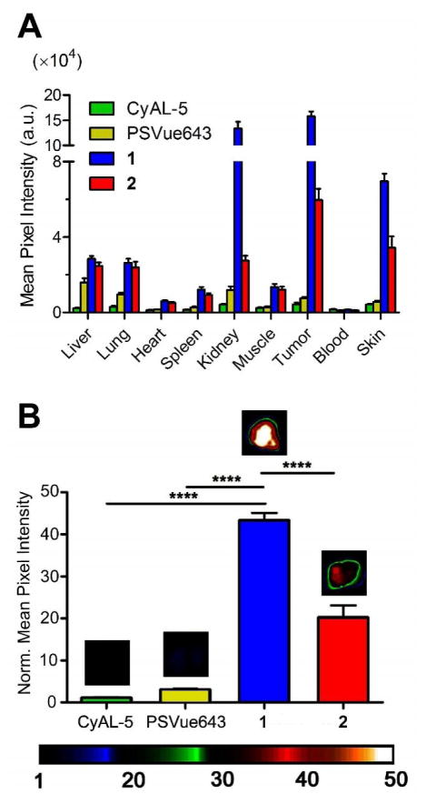
Figure 6.
Probe localization in a Lobund-Wistar rat subcutaneous prostate tumor model of cell death 24 h after probe injection. (A) Fluorescence biodistribution from excised organs. (B) Mean pixel intensities of excised tumors (normalized to CyAL-5 mean pixel intensity) showing high relative accumulation of Zn2TyrBDPAs. Error bars are standard error of the mean. N = 4, 4, 10, 6 for CyAL-5, PSVue643, 1, and 2, respectively. ****P ≤ 0.0001 Each cohort was given a tail vein injection of fluorescent probe (150 nmol) in water (1 % DMSO). The animals were euthanized 24 h later and biodistributions were determined by imaging the excised tissues using a planar fluorescence imaging station with a deep-red filter set (λex = 630 nm, λem = 700 nm).

