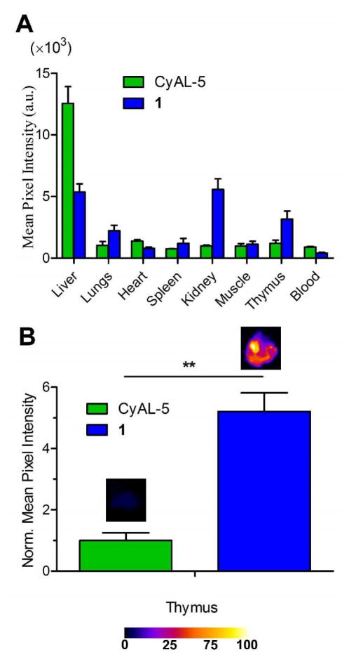
Figure 7.
Probe 1 localization in a thymus atrophy model of cell death in immunocompetent mice. (A) Biodistribution of probe 1 (blue) and untargeted CyAL-5 dye (green) in excised organs taken from mouse cohorts 3 h after intravenous injection of probe. Dexamethasone dosage was performed 24 h prior to probe injection. (B) Mean pixel intensities for probe fluorescence in the excised thymi. Error bars are the standard error of the mean. N = 3 for both cohorts. P values ≤ 0.01 (*), ≤ 0.001 (**) or ≤ 0.0001 (***) are considered statistically significant. SKH1 mice were given intraperitoneal injections of dexamethasone at a dose of 50 mg/kg. After 24 h, the imaging probe (10 nmol) was injected via the tail vain, and after 3 h mice were sacrificed and organs were imaged using a planar fluorescence imaging station.

