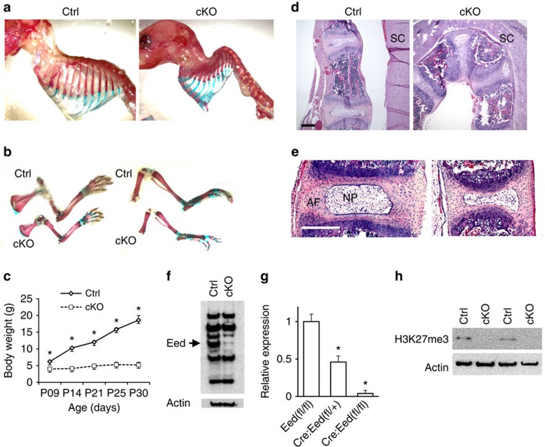
Figure 1. Conditional Eed deletion in chondrocytes causes skeletal defects.
(a,b) Whole-mount skeletal preparation of littermate control (Ctrl) and Eed cKO mice at P11. Both male and female cKO mice show severe kyphosis (Upper) and shortening of long bones of forelimbs (lower left) and hind limbs (lower right). (c) cKO mice show impaired growth. A majority of cKO mice dies by P21. Male and female mice were combined. n=3–6; *P<0.05 versus Ctrl, error bars show the s.e.m., unpaired Student's t-test was used. (d) Sagittal sections of thoracic vertebrae of mice at P28.5. SC, spinal cord. (e) Intervertebral discs are also affected. Eed cKO mice show reduced size of the annulus fibrosus (AF) and nucleus pulposus (NP). (f) Immunoblot analysis using protein lysate of primary rib chondrocytes cultured overnight shows an efficient reduction in the Eed protein in cKO chondrocytes. The arrow indicates the signal specific to Eed. (g) The Eed RNA expression level determined by qPCR in chondrocytes from indicated genotypes. (h) H3K27me3 is absent in primary rib chondrocytes of cKO mice (Supplementary Fig. 6; the uncropped blot result images). Scale bars, 200 μm.

