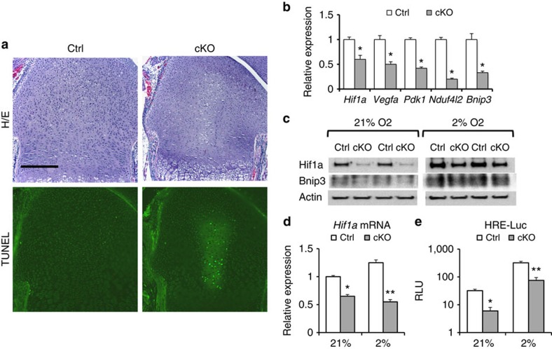
Figure 3. Eed deletion impairs hypoxic adaptation of chondrocytes.
(a) Haematoxylin/eosin (top) and TdT-mediated dUTP nick end labeling (TUNEL; bottom)-stained sections of the distal femoral growth pate of P1.5-old mice. Cell death is detected in the central area of the Eed cKO growth plate. (b) Expression levels of Hif1a and indicated Hif1a target genes were reduced in primary Eed cKO chondrocytes cultured in the normoxic condition. RNA expression levels were determined by qRT–PCR. Expression levels were normalized to Actb levels. n=4; * P<0.05 versus Ctrl. (c) Expression of Hif1a and Bnip3 is reduced in primary cKO rib chondrocytes both in normoxic (21% O2) and hypoxic (2% O2) conditions. (d) The Hif1a mRNA level is significantly reduced in cKO chondrocytes. Hif1a levels were normalized by 18S ribosomal RNA. n=4; *P<0.05, **P<0.01 versus Ctrl. (e) Hif1a activity, assessed by a Hif1a-responsive luciferase reporter (HRE-luc), was reduced in cKO in both normoxic and hypoxic conditions. n=6; *P<0.05, **P<0.01 versus Ctrl. Scale bars, 200 μm. Error bars show the s.e.m., unpaired Student's t-test was used.

