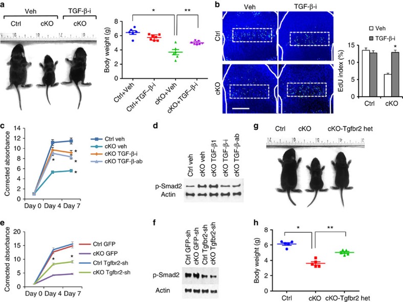
Figure 5. TGF-β inhibition rescues the growth defect of Eed cKO mice.
(a) The TGF-β receptor inhibitor, Ly364947, was injected daily into pregnant or nursing mothers from E14.5 through P7.5. Mice at P7.5 are shown. Body weight was measured at P7.5. n≥6; *P<0.05 versus Ctrl, **P<0.05 versus vehicle-treated cKO. (b) EdU staining of proximal tibial growth plates. The fraction of EdU-positive nuclei was calculated in proliferating columnar chondrocyte zone (dotted lines). White solid lines indicate the epiphyseal bone contour. n=3; *P<0.05 versus vehicle-treated cKO mice. Scale bars, 200 μm. (c) Cell proliferation assay on primary chondrocytes isolated from wild-type (Ctrl) and Eed cKO (cKO) mice, treated with dimethylsulphoxide (DMSO; Veh) or a TGF-β receptor inhibitor (TGF-β-i; Ly364947, 0.2 μM), TGF-β ligand-neutralizing antibody (TGF-β-ab; 5 μg ml−1) or TGF-β1 (20 ng ml−1). Treatment with Ly364947 and TGF-β-ab significantly ameliorated the proliferation defect of cKO cells. n=3; *P<0.01 and **P<0.05. (d) Treatment with TGF-β-i and TGF-β-ab decreases the p-Smad2 level. (e) Primary rib chondrocytes from Ctrl and cKO mice were infected with retroviruses expressing Tgfbr2 shRNA (Tgfbr2-sh) or eGFP (enhanced green fluorescent protein; GFP). Tgfbr2 knockdown using Tgfbr2-sh2 virus in cKO chondrocytes significantly increases proliferation. n=6; *P<0.05. (f) The p-Smad level is decreased in Tgfbr2 knockdown chondrocytes. (g) Tgfbr2 heterozygosity (cKO-Tgfbr2 het) significantly improves animal growth of Eed cKO mice. (h) Body weight of control (Ctrl), Eed cKO (cKO) and Eed cKO mice missing one allele of Tgfbr2 (cKO-Tgfbr2 het) was measured at P7.5. Mice with conditional Tgfbr2 heterozygous deletion alone (Col2-Cre:TgfbrloxP/+) show no noticeable skeletal abnormalities, as also previously described37. n≥5; *P<0.01 and **P<0.05. Error bars show the s.e.m., unpaired Student's t-test was used.

