
Figure 3.
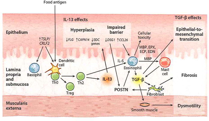
Pathophysiology of EoE. TSLP is released from the epithelium and activates basophils and food antigen–presenting dendritic cells to induce Th2 polarization of naive CD4+ T cells. These Th2 cells then secrete JL-13 that increases CCL26, CAPN14, and periostin (POSTN) expression and decreases DSG1, FLG, and DC gene expression in the epithelium. Decreased DSG1 level impairs barrier function, thereby forming a propagation loop by allowing further penetration of food antigen, and also leads to increased POSTN levels. The increased CCL26 and POSTN promote eosinophil recruitment from the bloodstream. The accumulating activated eosinophils cause epithelial cell cytotoxicity. TGF-β released by both eosinophils and mast cells increases POSTN expression and stimulates fibrotic response and smooth muscle dysmotility. Abbreviations: CAPN14, calpain 14; CCL26, chemokine (C-C motif) ligand 26; CRLF2, cytokine receptor-like factor 2; DSG1, desmoglein 1; ECP, eosinophil cationic protein; EDC, epidermal differentiation complex; EDN, eosinophil-derived neurotoxin; EoE, eosinophilic esophagitis; EPX, eosinophil peroxidase; FLG, filaggrin; IL, interleukin; MBP, major basic protein; TGF-β, transforming growth factor β; Th0, naive T helper; Th2, T helper type 2; Treg, regulatory T cell; TSLP, thymic stromal lymphopoietin.