
Fig. 2.
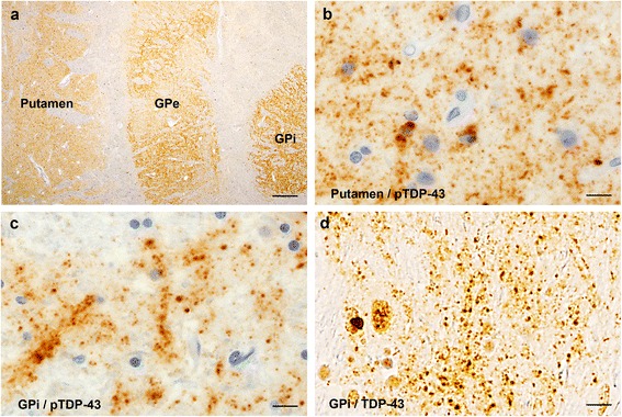
Dendrospinal pTDP-43 pathology in the neostriatum and globus pallidus. (a) Low-magnification view of the basal ganglia from a case of Type 2b delineates the putamen and globus pallidus with phosphorylated TDP-43 (pTDP-43) immunoreactivity. (b, c) High-magnification views of the putamen (b) and globus pallidus (c) show numerous pTDP-43-positive granular and dot-like structures. (d) Similar granular and dot-like structures in the globus pallidus are also visualized using phosphorylation-independent anti-TDP-43. Scale bars: a = 500 μm; b-d = 10 μm. GPe = external segment of globus pallidus; GPi = internal segment of globus pallidus