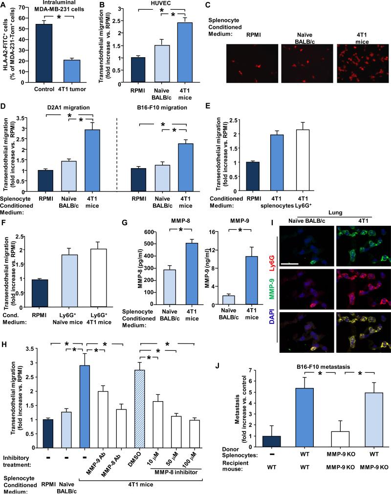
Figure 4. MMP-8 and MMP-9 facilitate transendothelial migration and metastasis.
(A) Intraluminal antibody staining of MDA-MB-231 cells injected i.v. into 4T1-mice (implanted with 4T1 cells 4 weeks earlier) or control naïve mice. Graph shows percent of MDA-MB-231-Tom+ cells found in the lung that also stained positive by i.v. injected anti-HLA-2-FITC antibodies. n ≥ 6 for each group. (B) Transendothelial migration of D2A1-Tom+ cells through HUVECs in the presence of the indicated conditioned media. Results present fold increase compared to control RPMI medium. At least 3 experiments were performed in duplicates. (C) Representative images of D2A1-Tom+ cells that migrated through HUVECs in the presence of the indicated conditioned media. (D) Transendothelial migration of D2A1-Tom+ cells or B16-F10-GFP+ cells through mouse endothelial, bEnd.3, cells in the presence of the indicated conditioned media. Results present fold increase compared to control RPMI medium. At least 3 experiments were performed in duplicates. (E) Transendothelial migration of D2A1 cells (fold increase compared to control RPMI medium) through mouse endothelial bEnd.3 cells in the presence of conditioned medium from unfractionated splenocytes or the Ly6G+ enriched subpopulation of splenocytes from 4T1 mice. The number of Ly6G+ cells used to prepare the conditioned medium was equivalent to the number of Ly6G+ cells in 1.5 ×10⁷ unfractionated splenocytes population.(F) Transendothelial migration of D2A1 cells (fold increase compared to control RPMI medium) in the presence of conditioned medium from the Ly6G+ enriched subpopulation of splenocytes from non-tumor-bearing or 4T1 mice. (G) MMP-8 and MMP-9 secretion by an equal number of splenocytes from naïve BALB/c or 4T1–bearing mice. (H) Transendothelial migration (fold increase compared to control RPMI) of D2A1cells in the presence of the indicated conditioned media, inhibitor or antibody against MMP-8 or antibody against MMP-9. At least 3 experiments were performed in duplicates. (I) Immunofluorescent staining of MMP-9 (green) and Ly6G (red) in lungs of naïve BALB/c and 4T1 mice (implanted with 4T1 cells 4 weeks earlier). Scale bar 25μm. (J) Adoptive transfer of splenocytes from WT or MMP-9 KO tumor-bearing mice (implanted with B16-F10-GCSF overexpressing cells 3 weeks earlier). Donor splenocytes were injected into WT or MMP9-KO recipient mice, as indicated. Experiment was terminated 2 weeks later, and pulmonary metastases of GFP-labelled B16-F10 cells were quantified. * indicates p< 0.05.

