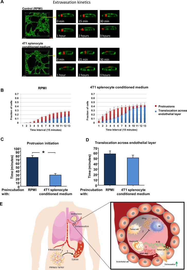
Figure 6. Visualization of protrusion and extravasation dynamics of tumor cells.
(A) Left - high resolution time-lapse confocal microscopy (20X) of an extravasating MDA-MB-231 cell (red) immediately after seeding in microvasculature network (HUVEC cells labeled in green). Right - confocal images taken at a single slice tracking a tumor cells as it transmigrates through the endothelium and into the 3D matrix over a period of 3 hours. (B) Protrusion formation (red) and extravasation (blue) rates of MDA-MB-231 cells in HUVEC vessels that had been pre-treated with 4T1 splenocyte conditioned medium or Control (RPMI). (C) Time required for protrusion formation (i.e. partial translocation across the lumen) and (D) time required for complete extravasation (i.e. the entire cell body has cleared the lumen) of MDA-MB-231 cells in HUVEC vessels that had been pre-treated with 4T1 splenocyte conditioned medium or Control (RPMI). Data were collected from >150 cells per condition, over 3 independent experiments. (E) Events leading to metastasis of circulating tumor cells: 1) Primary tumor perturbs distant organs, leading to neutrophil expansion and mobilization. 2) Neutrophils increase intraluminal survival of circulating tumor cells by inhibiting their clearance by NK cells. 3) Neutrophils increase extravasation of circulating tumor cells through secretion of IL-1β and MMPs. * indicates p< 0.05.

