Figure 1.
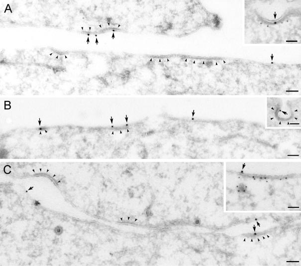
Recruitment of EGF-EGFR to clathrin-coated plasma membrane areas upon incubation with EGF on ice. Serum-starved Hep2 cells, incubated with EGF (60 ng/ml) on ice for 60 min, were prepared for immuno-EM and labeled for EGFR (A) or EGF (B and C). Labeling for EGFR and EGF (arrows) was found both at smooth and coated plasma membrane areas (coat indicated by arrowheads). That the coated areas represent clathrin-coated domains was confirmed by double-labeling for EGFR (large gold particles) and clathrin (small gold particles) shown as inset in A or EGF (large gold particles) and clathrin (small gold particles; inset in C). Although most of the EGF-EGFR positive coated areas were flat, some labeling localized to invaginated coated pits (inset in B). Bars, 100 nm.

