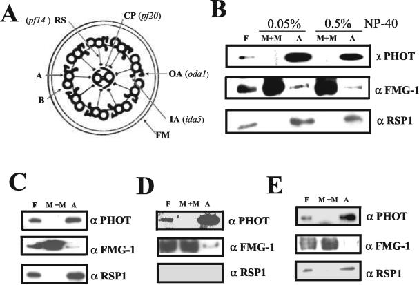
Figure 8.
Attempts to identify a phototropin interaction partner within the axonemal structure by the use of mutants. (A) Diagram of a cross section of a Chlamydomonas flagellum structure (adapted from Rosenbaum and Witman, 2002). The axonemal structures missing in individual mutants are indicated. Oda1 lacks the outer dynein arms (OA) (Kamiya, 1988; Takada et al., 2002); ida5 lacks most of the inner dynein arms (IA) (Kato et al., 1993; Kato-Minoura et al., 1997); pf14 has no radial spoke structures (RP) (Diener et al., 1990); pf20 lacks the whole central pair complex (CP) (Smith and Lefebvre, 1997). A and B represent the A- and B-tubules of outer doublet microtubules and FM the flagellar membrane. Flagella isolated from mutants oda1 (B), pf20 (C), pf14 (D), and ida5 (E) were separated into M+M and axoneme (A) fractions using 0.05% (oda1 mutant) and 0.5% NP-40 (oda1, ida5, pf14, and pf20 mutants) in the presence of 10 mM ATP in HMDEK buffer. The same amount of protein was loaded in each lane for Western blotting.

