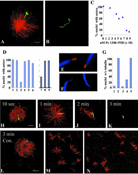
Figure 4.
C-terminal fragments of pericentrin disrupt aster formation and stability and γ tubulin assembly onto centrosomes in Xenopus mitotic extracts. (A and B) Mitotic asters assembled in the presence of equal amounts of pericentrin 1340–1920 (B) or heat inactivated (h.i.) 1340–1920 (A). (C) Mitotic aster assembly in the presence of increasing concentrations of pericentrin (Pc) 1340–1920. (D) Quantification of aster assembly in mitotic extracts in the presence of pericentrin domains. Amount of protein added per 10 μl of extract is indicated. (1, PBS; 2, 33 ng of 1–535; 3, 12 ng of 1340–1920 (p < 0.0001); 4, 12 ng of h.i. 1340–1920; 5, 10 ng of peri B1826–2117; 6, 10,000 ng of BSA). Quantification of aster assembly in interphase extracts (D7–9) by using a pericentrin domain (1618–1810) that inhibits aster assembly in mitotic extracts (7. p < 0.0001) and is inactivated by heat (8), but has no activity in interphase extracts in the same experiment (9). (E and F) γ Tubulin assembly onto nascent centrosomes in the presence of h.i. 1340–1920 (E) or 1340–1920 (F, p < 0.0001). (G) Quantification of γ tubulin assembly onto centrosomes in the absence of mitotic extract (1), in extracts with 1–595 (2), 1340–1920 (3, p < 0.0001), 1618–1810 (4, p < 0.0001), or heat inactivated 1618–1810 (5). For C, D, and G, 200 sperm nuclei were counted per bar or point. (H–K) Rapid disassembly of preassembled mitotic asters over time after addition of 1340–1920. (L) h.i. Pc 1340–1920 has no effect on preassembled asters. (M and N) Ran-mediated aster assembly in extracts in the presence of h.i 1618–1810 (M) or 1618–1810 (N). A–D, H–N, microtubules or γ tubulin, red; nuclei, green. Bar (A), 10 μm for A and B; in L, 10 μm for H–N.

