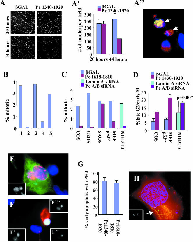
Figure 7.
Overexpression of GCP2/3 binding domain or silencing of Pc A/B induces cell cycle arrest and apoptosis at the G2/M phase of the cell cycle. (A) Cells expressing the GCP2/3 binding domain are lost through apoptosis. Low-magnification image of COS cells stained with DAPI showing cell loss when 1340–1920 is expressed for 44 h compared with nonexpressing cells (20 h) or β-galactosidase (βgal)–expressing cells. (A′) Quantification of cells in A (mean and SD, 10 fields). (A″) Image of COS cells from A stained for DNA (blue), 1340–1920 (red) and M30 cytodeath (green). Arrows, cells with both 1340–1920 and M30. (B) Mitotic index of U2OS cells expressing: 1, βgal; 2, 1618–1810 p < 0.001; 3, Pc B1826–2117 p = 0.437; 5, 1572–1816 p = 0.011; 6, 1572–1816m p = 0.226 (n = 1000 cells/bar at 40–44 h posttransfection). p values were calculated using the t-test relative to βgal controls). (C) Mitotic index in indicated cell types overexpression the indicated constructs or treated with siRNA. p values calculated as described above: COS, p < 0.001; U2OS, p < 0.001; SAOS, p = 0.479; Mefp53-/-, p = 0.001; NIH3T3 (siRNA), p = 0.0004. (D) Cells expressing GCP2/3 binding domain constructs or treated with Pc A/B siRNA have a greater proportion of late G2 cells. Shown are mean and SD of three experiments or p value based upon scoring from 1000 treated cells. Cells immunostained for overexpressed protein (green), phosphorylate histone H3 (PH3, red), and DAPI (blue). (E) Antephase cell overexpressing 1340–1920 (stains for phosphorylated histone H3 and does not show condensed chromatin). Inset, DAPI. (F) Apoptotic antephase cells expressing 1340–1920. M30 (red), phosphorylated histone H3 (green; inset, bottom right), DNA (blue; inset, above), overexpressed protein (inset, bottom left). (G) Graph showing that most early apoptotic cells expressing GCP2/3 binding domains stain for phosphorylated histone H3. Shown are mean, SD, n = 3 experiments. (H) Early apoptotic cell expressing 1340–1920 stained for centrosomes (5051, green), DNA (blue), HA pericentrin (red), M30, yellow. Inset 5051. Imaged as in Figure 5.

