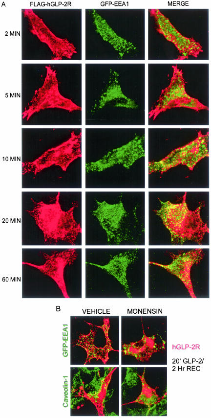
Figure 10.
hGLP-2R transiently colocalizes with early endosomes after agonist-induced internalization. (A) BHK cells cotransfected with FLAG-hGLP-2R and GFP-EEA1 were prelabeled on ice with anti-FLAG antibody and then treated with 10 nM hGLP-2 for 2–60 min at 37°C, as described in MATERIALS AND METHODS. (B) Transfected BHK cells were pretreated with vehicle or monensin for 30 min at 37°C, labeled with FLAG-antibody as in A, treated with 10 nM hGLP-2 for 20 min, washed, and allowed to recover in media alone for in the absence (vehicle) or presence of monensin for 2 h. After fixation and permeabilization, cells were stained with anti-caveolin-1-FITC and a Cy3-conjugated secondary antibody was used to visualize hGLP-2R/anti-FLAG antibody complexes. GFP-EEA1 was visualized directly. Confocal microscopy images are representative of two independent experiments.

