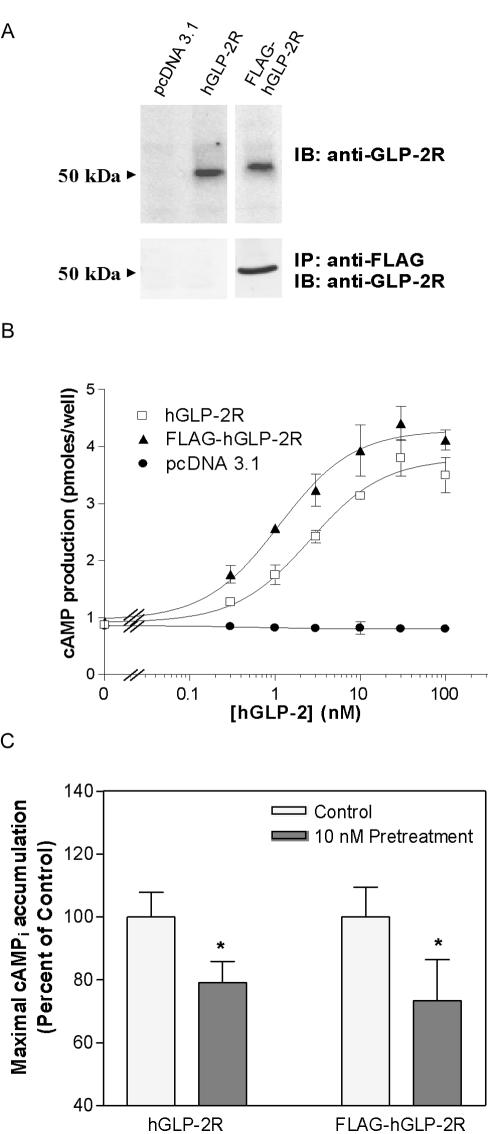
Figure 3.
An N-terminal FLAG-epitope does not alter hGLP-2-induced cAMP accumulation or desensitization of the hGLP-2R. (A) BHK cells were transiently transfected with either vector alone (pcDNA 3.1), untagged wild-type human GLP-2R (hGLP-2R), or FLAG-epitope–tagged human GLP-2R (FLAG-hGLP-2R). PNGase F-deglycosylated total cell extracts from BHK cells transiently transfected with the indicated constructs were analyzed by immunoblotting (IB) for the GLP-2R as described in MATERIALS AND METHODS. Alternatively, anti-FLAG antibody immunoprecipitates (IP) from the transfected cell extracts were subjected to deglycosylation before Western blot analysis to detect the GLP-2R. (B) Receptor activation was assessed by monitoring cellular cAMP production in response to hGLP-2 in transiently transfected BHK cells. Data were fit to a sigmoidal dose-response curve by using GraphPad Prism software. (C) DLD-1 cells transiently transfected with either the hGLP-2R or the FLAG-hGLP-2R constructs, were pretreated with vehicle or 10 nM hGLP-2, allowed to recover for 30 min, and then rechallenged with 100 nM hGLP-2 to determine maximal agonist-induced cAMPi accumulation. Data (means ± SD from triplicate values) were normalized and compared with control (*p < 0.05) as described in Figure 1.

