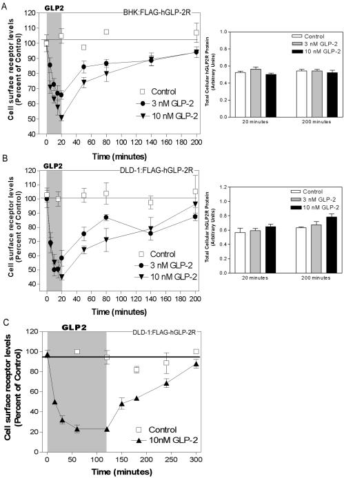
Figure 4.
Rapid agonist-induced hGLP-2R internalization is followed by slow cell surface receptor reappearance. BHK (A) or DLD-1 (B and C) cells transiently transfected with FLAG-hGLP-2R were treated with vehicle (control), 3 nM, or 10 nM hGLP-2 for either up to 20 min (A and B) or 2 h (C) to assess hGLP-2R internalization. After the hGLP-2 treatment, cells were washed briefly to remove the agonist and incubated for the indicated times in media with serum to assess hGLP-2R reappearance on the cell surface (left). Cell surface receptor levels were measured using an enzyme-linked immunoassay as described in MATERIALS AND METHODS. Data (means ± SD of triplicate values) are expressed as percentage of the cell surface receptor level at time 0. Similar results were obtained in two independent experiments for each cell type. Total cellular levels of FLAG-hGLP-2R in BHK cells (A, right) and DLD-1 cells (B, right) at the end of the internalization (20 min), and washout (200-min) periods were measured as indicated above, after permeabilization of the cells with 0.2% Triton X-100. Data are means ± SD of triplicate values (n = 2 independent experiments for each cell line).

