Figure 2.

(A and B) The pretreatment angiogram.
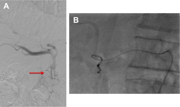
Notes: (A) This shows a very thin retroduodenal hepatic artery (arrow). (B) Superselective catheterization of the retroduodenal artery and embolization with microcoils.

(A and B) The pretreatment angiogram.
Notes: (A) This shows a very thin retroduodenal hepatic artery (arrow). (B) Superselective catheterization of the retroduodenal artery and embolization with microcoils.