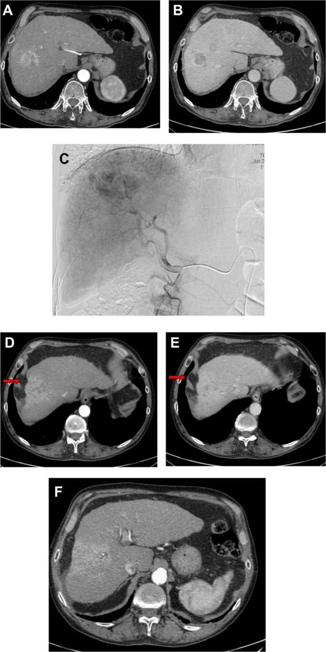
Figure 4.

(A–F) Bilobar HCC of the right hepatic lobe (V–VIII segments).
Notes: (A) The pretreatment CT shows hypervascularization of the larger lesion in the arterial phase and (B) the hypoattenuation of both nodules in the portal-venous phase. (C) The pretreatment angiogram confirms the large HCC nodule of the dome of the liver. CT study performed 8 months after treatment shows the complete devascularization of the lesions. Note the capsular retraction of the treated segment as a consequence of hepatic fibrosis (arrows) and the transient perfusion abnormalities in the treated area, persistent in both (D) the arterial and (E) the portal-venous phase which is, however, not a recurrent tumor. (F) Of note, the compensatory hypertrophy of the left lobe.
Abbreviations: HCC, hepatocellular carcinoma; CT, computed tomography.
