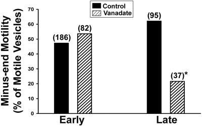
Figure 6.
Directional motility of early and late endocytic vesicles. Polarity-marked MTs were prepared and bound to the inner surface of a glass microscopy chamber. Early or late endocytic vesicles were then perfused into the chamber and bound to MTs. The bars indicate the percentage of early (left) and late (right) endocytic vesicles that moved toward the minus-end of MTs after addition of 50 μM ATP in the presence or absence of 5 μM vanadate. The total number of motile vesicles examined is shown in parentheses. *p < 0.002 compared with control late vesicles.

