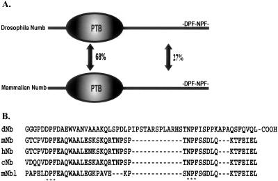
Figure 1.
Structure of Numb adaptor protein. (A) Schematic representation of Drosophila and mammalian Numb proteins. PTB-phosphotyrosine binding domain. (B) Multiple sequence alignment of Numb carboxy-terminal sequences in Drosophila (dNb), mouse (mNb), human (hNb), chicken (cNb), and mouse Numblike (mNbl). Asterisks mark the conserved DPF and NPF endocytic motifs.

