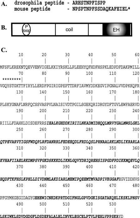
Figure 2.
Structure of the EHD/Rme-1 family of EH-domain–containing proteins. (A) Amino acid sequence of peptides used to screen expression libraries. Peptides were coupled to biotin at the amino terminus. (B) Schematic representation of domain organization of EHD protein family. P-loop, phosphate-binding motif; coil, coiled-coil domain; EH, Eps15-Homology domain. (C) Primary amino acid sequence of Drosophila EHD orthologue (accession no. AF473822). Asterisks indicate the p-loop consensus sequence. The coiled-coil domain is denoted by bold italic type, and the EH-domain is indicated by boldface type.

