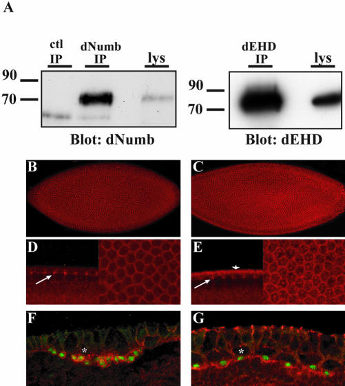
Figure 3.
Expression pattern of dNumb and dEHD during embryogenesis. (A) Whole flies were homogenized in lysis buffer, and immunoprecipitations with either affinity-purified anti-dNumb (left panel) or anti-dEHD (right panel) were performed. Western blotting revealed a single band in both immunoprecipitation and lysate (lys) lanes corresponding to the expected molecular weight of the dNumb or dEHD protein. (B--E) Whole-mount immunostaining of stage 4–5 Drosophila embryos during cellularization. dNumb expression (B) is found ubiquitously in all cells. dEHD expression (C) is similarly enriched at cell borders but is also found within large vesicular like structures. Lateral and face views of B and C at higher magnification demonstrating accumulation of dNumb (D) and dEHD (E) at the progressing furrow channel (arrows, left panel) and enrichment at apico-lateral cell borders (right panels). Apical staining of dEHD (arrowhead, E) corresponds to apical membrane or apical cytoplasm. (F and G) Lateral view of whole-mount immunostaining of stage 8–9 embryos double-stained with α-prospero (F and G, green) and α-dNumb (F, red), or α-dEHD (G, red). Asterisks demark apical neuroblast. Both dNumb and dEHD are coexpressed in Prospero-positive ganglion mother cells (GMC).

