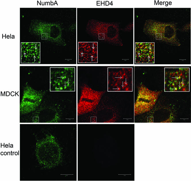
Figure 6.
Endogenous Numb and EHD4 colocalize in intracellular vesicles. HeLa (top and bottom panels) and MDCK (middle panels) cells were costained with rabbit anti-NbA plus goat anti-rabbit Fab (green), followed by rabbit anti-EHD4 (red) as described in MATERIALS AND METHODS. The bottom HeLa cell image is a representative cell taken from a control coverslip that was treated identically to the HeLa and MDCK cells shown above it except that no anti-EHD4 was added. The parameters used for image acquisition were the same. Confocal sections taken through the middle region of the cell are presented. The red and green images were merged to show regions of colocalization of mNumb and mEHD4 (yellow). Scale bar, 10 μm.

