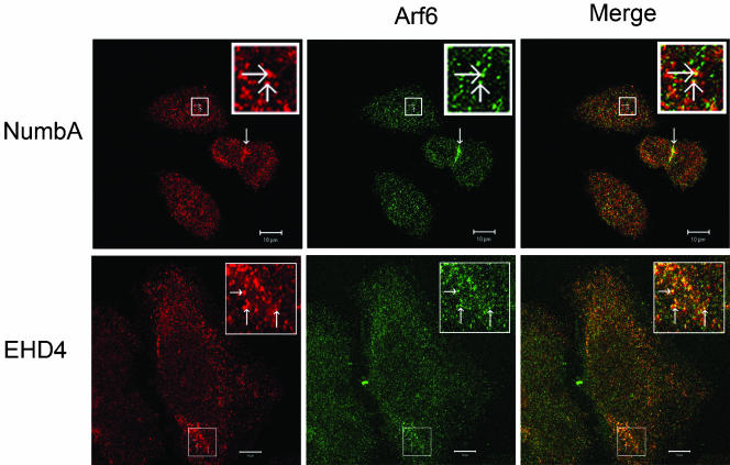
Figure 7.
Endogenous Numb and EHD4 colocalize with Arf6. HeLa cells were fixed and processed for immunofluorescence localization of endogenous Numb (top panel, red) or EHD4 (bottom panel, red), and Arf6 (green). The green and red images were merged to demonstrate colocalization of Arf6 and Numb or EHD4 (yellow). Numb and Arf6 were enriched at what appears to be the cleavage furrow (thickened arrow; top panel). Boxed regions have been digitally enlarged and represented as inserts in order to show more clearly examples of colabeling (arrows). Scale bar, 10 μm.

