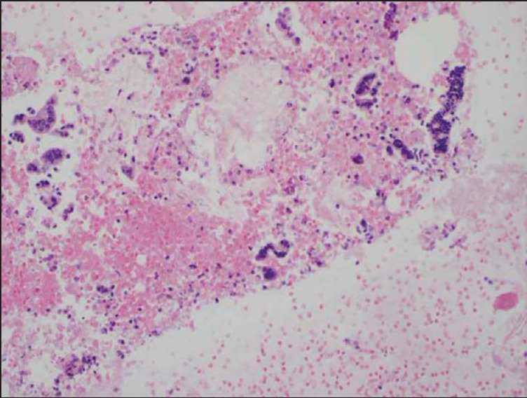
Figure 4.

Cell block with diagnostic material without a tissue core. Hematoxylin and eosin (20×) stain of a cell block preparation from a case using the EchoTip® needle. The photomicrograph shows malignant-appearing strips of columnar cells consistent with adenocarcinoma. It is to be noted that the cell clusters are not present in a true tissue core and are instead dispersed within a red blood cell background
