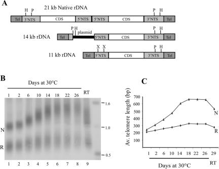
Figure 1.
Growth of rDNA telomeres. (A) Organization of native and recombinant rDNA molecules. Coding sequences (CDS) are represented as open boxes, 5′ and 3′ NTS are light-shaded boxes, and telomeres are dark boxes. HindIII (H), PstI (P), and XbaI (X) sites are marked. (B and C) Differential growth of 14-kb rDNA telomeres with natural and recombinant subtelomeric regions. (B) Native (N) and recombinant (R) telomeric restriction fragments from the 14-kb rDNA were detected by Southern hybridization with probe for telomeric DNA. Cells were grown at 30°C for 1–26 d then at room temperature (RT) for 3 d. (C) Average length of the telomeric tract from the native and recombinant telomere is plotted against time in culture.

