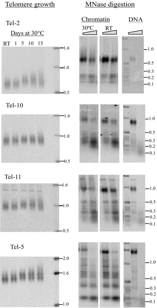
Figure 6.
Telomere growth and chromatin structure at non-rDNA telomeres. Left, telomeric restriction fragments from different chromosome ends after growing strain CU428 at room temperature (RT) or in continuous culture at 30°C. Middle, corresponding MNase digests of subtelomeric chromatin. Macronuclei were isolated from cells grown at RT or for 30°C for 15 d. Digestion was with 30 U of MNase/mg chromatin for 4 and 6 min. Right, deproteinized DNA controls digested with 8 U of MNase/mg DNA for 1 and 2 min. Purified MNase digestion products were restriction digested and analyzed by Southern hybridization with a probe that hybridized adjacent to the restriction site.

