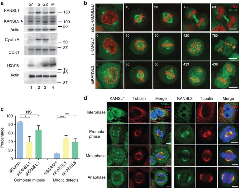
Figure 1. KANSL1 and KANSL3 localize to spindle poles in mitosis and their depletion leads to mitotic defects.
(a) NSL complex members KANSL1 and KANSL3 are expressed throughout the cell cycle in HeLa cells. Synchronization was confirmed by western blotting of various cell cycle markers, as indicated. Molecular weight markers are indicated on the right and actin was used as a loading control. (b) Cells exhibit mitotic defects on knockdown of NSL complex members KANSL1 and KANSL3. Representative still images from the live-cell analysis show dividing control (siSCRAMBLED) and KANSL1- and KANSL3-silenced H2B-mCherry/a-tubulin-GFP HeLa cells. Typical phenotypes exhibited on knockdown of KANSL1 and KANSL3 include misaligned chromosomes persistently attached to spindle poles, a prolonged metaphase delay and mitotic catastrophe (note membrane blebbing in the final panel). Time in minutes is indicated in the upper left corners. Scale bars, 10 μm. (c) Left side: quantification of the percentage of cells that complete mitosis over a 24-h time frame. All other cells remained arrested in prometaphase or had died by the end of the observation period. Right side: quantification of the percentage of mitotic cells exhibiting the defects of misaligned chromosomes, multipolar spindles, lagging chromosomes or mitotic catastrophe. Error bars, s.e.m. *P<0.05, **P<0.01, NS, non significant, according to unpaired one-tailed t-test. (d) Immunofluorescence of KANSL1 or KANSL3 proteins (green) through the cell cycle. Tubulin is in red and DNA is in blue. Scale bars, 5 μm. KANSL1 and KANSL3 localize to spindle poles in mitosis.

