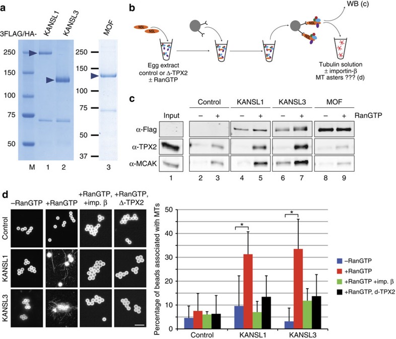
Figure 2. KANSL1 and KANSL3 promote microtubule assembly in a RanGTP-dependent manner in Xenopus egg extracts.
(a) Coomassie blue-stained gel of purified recombinant Drosophila KANSL1, KANSL3 and MOF tagged with 3FLAG/HA. (b) Schematic representation of the experimental design. KANSL1 or KANSL3 was incubated in egg extract±RanGTP and retrieved on magnetic beads coupled with anti-HA antibodies. Beads were analysed by western blot or incubated in pure tubulin to monitor microtubule assembly. (c) Western blots of KANSL1, KANSL3 and MOF beads retrieved from M-phase Xenopus egg extracts in the presence (+) or absence (−) of RanGTP. The control was performed in parallel with egg extract in the absence of recombinant proteins. Anti-Flag antibodies were used to visualize KANSL1, KANSL3 and MOF. KANSL1 and KANSL3 interact with TPX2 and MCAK in a RanGTP-dependent manner. (d) Microtubule assembly activity in pure tubulin. The KANSL beads retrieved from M-phase Xenopus egg extracts in the presence (red bars) or absence (blue bars) of RanGTP were incubated in 20 μM pure tubulin. Green bars correspond to beads retrieved from an extract containing RanGTP and then incubated with importin-β and pure tubulin. Black bars represent beads retrieved from a xTPX2-depleted D-TPX2 egg extract containing RanGTP. Beads were spun down on coverslips and processed for immunofluorescence to visualize microtubules. Representative images of beads are shown on the left. Beads are autofluorescent. Scale bar, 5 μm. The graph on the right shows the percentage of beads interacting with microtubules in each condition. KANSL1- and KANSL3-coated beads induce the assembly of microtubules when retrieved from an egg extract containing RanGTP. Data are from three independent experiments. Error bars, s.d. *P<0.05 (unpaired t-test), NS, non significant.

