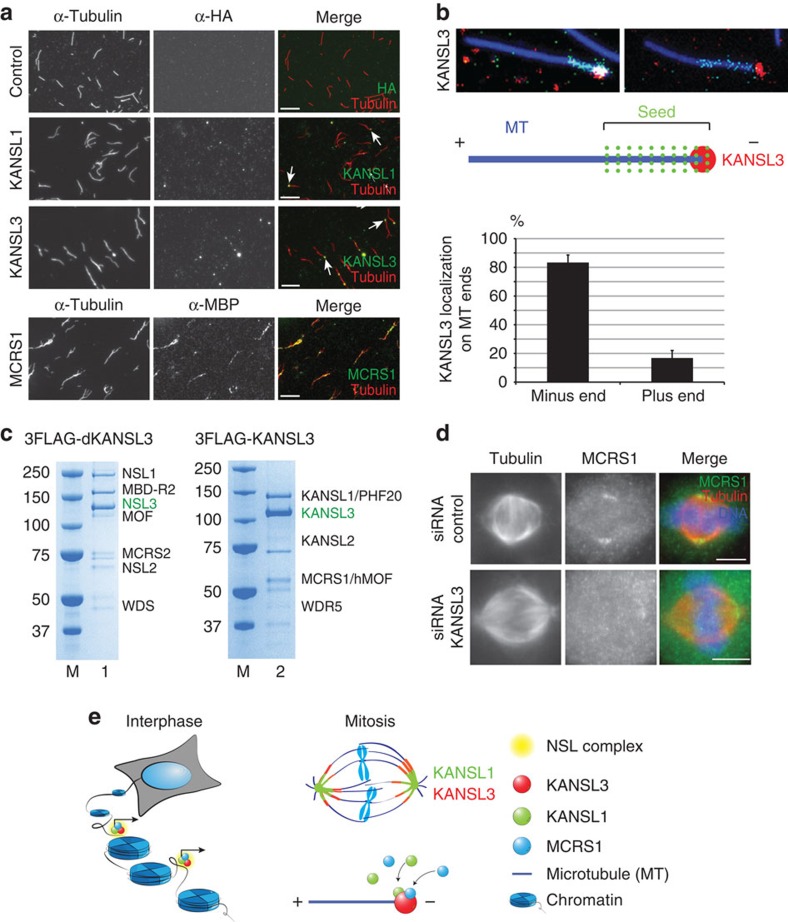
Figure 4. KANSL3 is a microtubule minus-end-binding protein.
(a) KANSL1 and KANSL3 localization to microtubule ends in vitro. Taxol-stabilized microtubules were incubated with recombinant KANSL1, KANSL3, xMCRS1 or buffer (control), as indicated. Microtubules were spun down on coverslips and processed for immunofluorescence with anti-tubulin (red) and either anti-HA (for KANSL proteins) or anti-MBP (for MCRS1) antibodies (green). Arrows indicate KANSL1 and KANSL3 accumulation at microtubule ends. Scale bar, 5 μm. (b) KANSL3 binds to the minus end of polarity-marked microtubules in vitro. Polarity-marked microtubules were incubated with KANSL3, spun down on coverslips and processed for immunofluorescence. The bright microtubule seed at the minus end is in green, tubulin is in blue and KANSL3 in red, as shown in the drawing. KANSL3 associates with the microtubule minus end. Bottom: quantification of the microtubule minus end localization of KANSL3. The percentage of plus-end and minus-end signal for KANSL3 is shown. KANSL3 binds preferentially to minus ends (81.3%) versus plus ends (18.7%). n=3. More than 250 microtubules were analysed. Error bars, s.d. (c) KANSL3 nucleates a complete NSL complex in vitro. All seven members of the NSL complex were expressed in SF21 cells as untagged proteins except KANSL3 that was tagged with 3FLAG. Flag pull-down from the cellular extract retrieved a complete heptameric NSL complex. This indicates that KANSL3 is a structurally central component of the NSL complex, both in the context of the Drosophila and human proteins. (d) KANSL3 silencing in HeLa cells results in the loss of MCRS1 localization to spindle poles. Representative immunofluorescence pictures of control or KANSL3-silenced HeLa cells, as indicated. Tubulin is in red, MCRS1 in green and DNA in blue. Scale bar, 5 μm. (e) Summary. In interphase, KANSL1 and KANSL3 proteins are chromatin bound and regulate expression of housekeeping genes. During mitosis, these proteins relocate to the mitotic spindle and are important for cell division. Moreover, KANSL3 binds directly to microtubule minus ends in vitro and localizes to K-fibre microtubule minus ends in the dividing cell. Thus, KANSL proteins are able to adopt distinct tasks in different phases of cell cycle to ensure cellular homeostasis.

