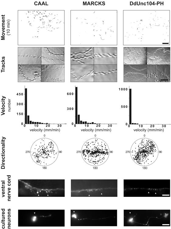
Figure 7.
Substitutions of the UNC-104 PH domain with other lipid-binding modules only weakly or fails to rescue the unc-104 phenotype. The movement of the transgenic worms (scale bar, 4.8 mm), agar tracks (scale bar, 0.13 mm), worm velocities, directional persistence, and analysis of synaptotagmin staining in the ventral nerve cord (scale bar, 10 μm) and in cultured neurons (scale bar, 10 μm) were performed as described in the legends to Figures 2, 3, 4. Cell body staining is indicated by the arrowheads. See text for description of phenotypes.

