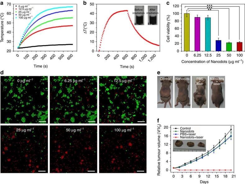
Figure 5. Photothermal therapy of Fe-CPNDs.
(a) Temperature elevation of the water and Fe-CPNDs solutions of various concentrations (Fe content: 0–100 μg ml−1) over a 10-min exposure to 808-nm NIR light (1.3 W cm−2). The temperatures were measured every 10 s using a thermometer. (b) The photothermal response of the 100 μg ml−1 (Fe content) Fe-CPND aqueous solution when exposed to 808-nm NIR light (1.3 W cm−2) for 600 s. The laser was then turned off. The inset shows the digital photograph of the dispersion of Fe-CPNDs in water before and after laser irradiation. (c) In vitro cell viabilities of SW620 cells incubated with various concentrations of Fe-CPNDs (Fe content: 0–100 μg ml−1) under 808-nm NIR laser irradiation (1.3 W cm−2, 6 min). The error bars indicate the s.d. (n=5, **P<0.01 or ***P<0.001 from an analysis of variance with Tukey's post-test). (d) Confocal fluorescence images of SW620 cells after incubation with various concentrations of Fe-CPNDs (Fe content: 0–100 μg ml−1) under 808-nm NIR laser irradiation for 6 min. The cells were co-stained with calcein acetoxymethyl ester (green, living cells) and propidium iodide (red, dead cells). Scale bar, 200 μm. (e) Digital photographs of the mice collected from different groups at the end of intravenous treatments (day 20). (f) Corresponding tumour growth curves of different groups of mice after intravenous treatments. Error bars indicate the s.d. (n=4). The inset shows the digital photographs of tumours collected from different groups of mice at the end of intravenous treatments (day 20).

