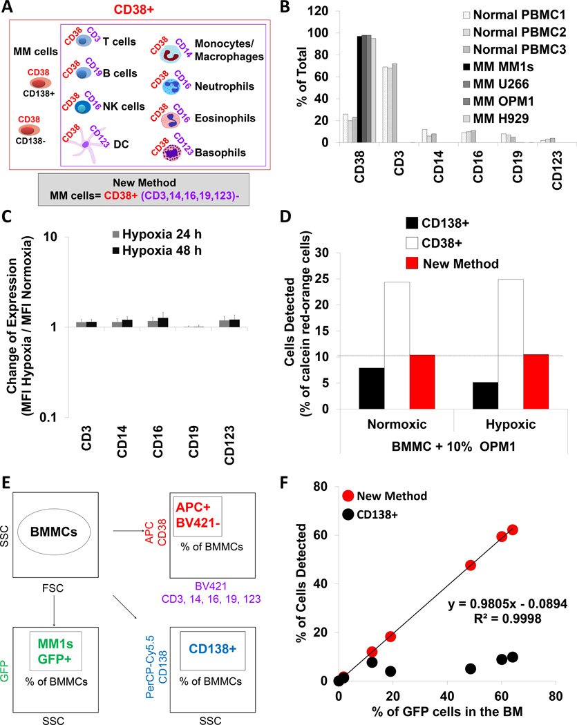
Figure 2. Development and validation of the new strategy to detect MM cells.
(A) The methodology behind the new strategy to detect multiple myeloma (MM) cells. (B) The percentage of cells positive for CD38, CD3, CD14, CD16, CD19 and CD123 in the normal PBMCs from 3 healthy donors and 4 MM cell lines detected by flow cytometry. (C) Expression of CD3, CD14, CD16, CD19 and CD123 in MM cells exposed to hypoxia (1% O2) for 24 and 48 h, and normalized to normoxia (21% O2), shown as a mean ± s.d. from 5 MM cell lines. (D) Detection of normoxic and hypoxic calcein red-orange+ OPM1 cells spiked into BMMCs detected by flow cytometry using CD138, CD38 and the new method. (E) The experimental approach of detecting MM cells in the BMMCs from MM-bearing mice using three strategies: GFP+, CD138+ and the new method. (F) Percentage of MM cells identified by CD138+ or the new method and plotted against the percentage of GFP+ cells in the BM from mice with different tumour size. GFP - green fluorescent protein; DC – dendritic cells; BMMCs – bone marrow mononuclear cells; PBMCs – peripheral blood mononuclear cells; SSC – side scatter; FSC – forward scatter.

